Renal Trauma
ASST
Organ injury scale for renal trauma
|
Grade |
Injury |
|
I |
Non-expanding
subcapsular haematoma without parenchymal laceration Contusion
with microscopic or gross haematuria and normal
investigations |
|
II |
Laceration
<1cm parenchymal depth without urinary extravasation
|
|
III |
Laceration
>1cm without collecting system rupture |
|
IV |
Laceration
extending into the collecting system with urinary
extravasation; injury to main renal vessel with
contained haemorrhage |
|
V |
Shattered
kidney; avulsion of renal hilum with devascularization |
Evaluation
1. Haematuria is the standard marker for renal injury; perform
in all patients
- bladder and ureter may also show haematuria.
- may not occur with injuries to the proximal renal
vasculature.
2. Blunt trauma
- beware deceleration injury; renal pelvis / uretopelvic jx
injury
- look for flank ecchymosis, lower rib fractures, transverse
process #
3. Penetrating trauma
- identify wounds with radio-opaque markers to help imaging
- high risk in posterior wounds; anterior usually hit other
structures first.
4. Indications for renal imaging:
- blunt trauma and gross haematura
- blunt trauma, haematuria, shock
- major accleration or deceleration injury
- any haematuria after penetrating injury
- associated injuries and physical signs
5. Imaging
- CT: best choice
- arteriographic phase for hulum injuries, blush.
- renal vein injuries identified by haematoma medial to
kidney.
- USS: blood in Gerota will not show in FAST not effective for
kidneys
- IVU replaced by CT, but may do a well timed IVU prior to
theatre to confirm contralateral kidney fx(?)
- arteriography used for intervention.
6. Instability
- no need for imaging; laparotomy.
7. Indications for renal exploration
- vast majority can be managed non-operatively
- absolute = peristent life threatening bleeding
- relative = devitalized parenchyma >50%, urinary
extravasation not resolving (25% or so) - usually stented
though if possible; arterial thrombosis; penetrating injuries
Surgical Management
1. Retroperitoneal injuries
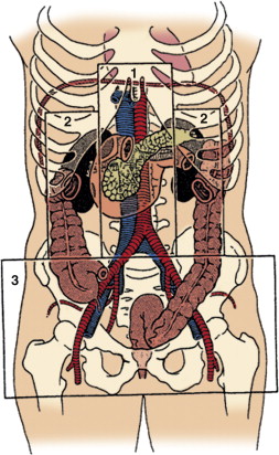
- need specialist input, often need patch repair / grafts
- no need for proximal control in general; sdisplace small
bowel along root of mesentery and laterally to right.
- take down the ligament of treiz
Non-Operative Management
Recent evidence suggests that bed rest can be
avoided unless bleeding appreciably increases or resumes after
ambulation.
Complications
- other complications include delayed bleeding, arterial
pseudoaneurysm, urinary fistula, hydronephrosis
- hypertension; can be transitory
Rest of Jerome's Notes
· Can affect
— Parenchyma
— Capsule
— Pedicle
· Either Penetrating
or blunt. Blunt most common.
· Risk of renal
trauma is greater in children
· Mechanism of
injury, haematuria (gross or microscopic) or associated injuries
(lower rib #) should raise the possibility of renal trauma.
· Haematuria is the
most sensitive indicator of injury but does not predict
severity.
· Radiographic
investigation is required in:
· Macroscopic
haematuria
· Microscopic
haematuria and episode of SBP<90
· Microscopic
haematuria and SBP>90 with suspicious mechanism of injury, or
physical exam (lower rib # or flank bruise).
CT is the best
study with IV contrast. An arterial phase and a 10min delayed
scan to detect collecting system injury.
Single shot IVP
· 2ml/Kg of
IV contrast is given and then a KUB is taken 10 minutes later.
· Used for unstable
patient who is to proceed to theatre for immediate exploration.
Its major value is in assuring that contralateral
kidney is functional.
Penetrating
· Classification
— Perforating
— Lacerating
— Explosive
· Often complicated
by perforation of other structures
— (90% gunshot, 60%
stab)
— R side
Liver/colon (60%)
— L side
Spleen/stomach/pancreas
(60%)
— Chest (20%)
Clinical
· Shock 30%
· Associated injuries
70%
· Gross haematuria
60%
· Renal or clot colic
— Micro haematuria
³20%
v NB absence of
blood does NOT exclude injury
Blunt
Classification
Mechanism
· Direct
· Indirect
Severity
· Major
— pedicle rupture,
pelvic tears, lacerations
· Minor
— Contusions, small
subcapsular haematomas
Aetiology
· Acceleration/deceleration
— Intimal tear ® renal artery
thrombosis
— Artery 70%
— Vein 20%
— Both 10%
— Mortality » 50%
Clinical
· Shock 5%
· Associated injuries
15%
· Gross haematuria
· Micro haematuria
Ix
· Urinalysis
· IVP single shot
— Extravasation /
non visualisation
· CT
Rx
Haemodynamically
stable patients can most often be managed non-operatively
Grade I-IV injuries
often managed non-operatively.
Indications for
exploration:
· Unstable
with renal haemorrhage
· Penetrating
injury
· Expanding
or pulsatile retroperitoneal haematoma at laparotomy
· Grade V
injuries
Operative
principles
· Mobilize colon to
expose Gerota’s fascia
· Manual compression
of hilum followed by clamping – about 45minutes of warm
ischaemic time is tolerated.
· Open Gerota’s
fascia
· Nephrectomy: Grade
V injuries, continuing haemodynamic instability.
· Repair for all
other injuries if possible: haemostasis, debridement, collecting
system repair & pledgeted bolstered closure of the renal
capsule.
Complications
Immediate: bleeding
– leave a closed suction drain
Early: Ileus,
infection, pneumonia, secondary bleeding, urinoma – can usually
be managed expectantly and some cases will require drainage,
stenting or later exploration
Late:
AV
malformation – usually heralded by bleeding at 3-4 weeks (treat
with selective embolization)
Hypertension <2% (within first several month) from either renal
artery stenosis, renal parenchyma compression, or AVM (chronic renal
ischaemia leads to rennin release).
Ureter
Classification
Iatrogenic
Penetrating
· Associated with
other injuries
— Small bowel 80%
— Colon 60%
— IVC 20%
GSW much more
common than SW
Blunt
· < 10% of
injuries
Clinical
· Abdo pain
· Fever
· Haematuria
· Delayed
presentation often occurs with fever, flank pain, urioma, renail
failure, ileus, urinary fistula
Ix
· IVP
· Retrograde
pyelogram
Rx
· If a
ureteric injury is suspected during laparotomy then inject indigo carmine and
look for leakage of blue dye.
Damage control:
· Only perform a
definitive reconstruction if the patient is stable
· Otherwise ligate
ureter, drain retroperitoneum and bladder and place a perc
nephrostomy
Definitive
repair
· Lower
— Reimplantation –
if the ureter is too short use a vesio-psoas
hitch or Boari flap
· Mid
— Primary anastomosis
– spatulated end-to-end
— Transuretoureterostomy
— ± renal
mobilisation
· Proximal
— Primary
anastomosis
— ± pyeloplasty and
renal mobilization
Or complex injury
kidney Autotransplant
Ileal interposition
ureterocalicostomy
Psoas Hitch
Apex of bladder sutured to the ipsilateral psoas tendon;
contralateral superior vesicle pedicle divided to improve
bladder mobility; care not to trap genitofemoral nerve.
Principles of
definitive repair are
tension
free
mucosa-to
mucosa
spatulated (cut the tube longitunally so allowing greater
circumference to join)
stented
drained
using
absorbable suture.
Bladder
Causes:
Iatrogenic
Penetrating injury
Blunt trauma
· 75% of bladder
injuries
· 10% of pts with
pelvic #
· Direct blow, full
bladder
Signs
-
Haematuria
-
Suprapubic pain
-
Inability to void
-
Incomplete recovery of catheter irrigation
Rupture
· Extraperitoneal:
Location: bladder base
and parasymphyseal area
from pelvic symphseal #
or ramus #
Treated with IDC for 10
days then reimaging to confirm healing
· Intraperitoneal:
Locations: Dome of bladder
From full bladder and direct blow
Laparotomy for repair
Ix
· Haematuria
· Exclude urethral
injury
· Retrograde cystourethrography (If suspected upper
tract injury, do IVP first):
Fill bladder with 400ml of 50% diluted contrast
AP/Oblique/Lateral views + post-voiding film
Sunburst pattern for intraperitoneal rup: contrast in SB
loop, gutters, organs
Contrast leaking adjacent to symphysis for
extraperiotneal injury
· CT cystography
Rx
· IDUC/SPC
· Primary repair
Urethra
Common in Male than
Female due to length of urethra
Mechanism
-
Crushing/deceleration injury
-
Staddle
-
Pelvic #
§ Commonly rupture at
prostatomembraneous uethra
-
During Sexual intercourse
Signs
-
Haematuria
-
Bruise or Swelling penis or scrotum
-
PR: high riding prostate
-
Inability to void
-
Inability to pass catheter
Rx
· Incomplete transaction (Regardless site)
Stenting across the
defect
or
Diversion with
suprapublic cystostomy
· Complete transaction
Always needs suprapubic cystostomy
Endoscopic bridging IDC
Complications
· Structure
· Incontinence
· Impotence
Posterior
Classification
· Urogenital
diaphragm
· Ext sphincter
· Prostatic
Associations
· 5% of pts with
pelvic #
· 50% with disruption
of pelvic rim
Ix
· Rectal
— Boggy mass
— Mobile prostate
· Retrograde
urethrogram prior to insert IDC
Rx
· Controversial
— Pass/not pass
IDUC
— SPC
— Primary repair
Anterior
Classification
· Bulbar
· Pendulous
Aetiology
· Iatrogenic
· Straddle
· (gunshot !)
Pathology
· Extravasation
within Colles’ fascia
Clinical
· Pain
· Swelling
· Echymosis
· Blood urethral
meatus
· Inability to void
Trauma 34
Ix
· Retrograde
urethrogram
Rx
· Partial seperation
— Silastic IDC,
antibiotics
· Full seperation
— Perinieal
incision, drainage and primary repair